


RETINA
WHAT IS RETINA?
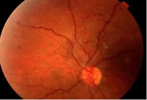
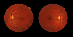
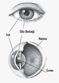
The retina is the layer that contains light-sensitive cells and nerve fibers that enable us to see. The network layer we call retina covers the back inner wall of the eyeball, just like a wallpaper. The retina consists of millions of vision cells and the nerve cells to which they are connected. The extensions of these nerve cells (about 1.5 million) come together to form the optic nerve. The vessels that feed these cells are also located within the retina. The specialized area in the retina that provides central vision and focuses the light is called the macula (yellow spot).
Defect in the retina causes the image not to be formed, and defect in the optic nerve causes the image not to reach the brain or to reach incompletely. The person loses his ability to see.
Bodrum Eye Retinal Diseases, diagnosis and treatment of all diseases related to the Retina and Intraocular Gel (vitreous) are carried out using the latest technology developed in the world. The two most important diseases that threaten the retina are diabetes and hypertension.
- Symptoms of Retinal Diseases
- Sudden or gradual loss of vision,
- distorted vision
- flashes of light,
- flies flying,
- Objects moving in front of the eye,
- obstruction of vision,
- Temporary and short-term vision loss,
- Dark areas in the field of vision.
How is it diagnosed?
Depending on the type of retinal disorder, medical treatment, laser treatment and surgical treatment may be required.
YELLOW SPOT DISEASE (Macular Degeneration)
The macula is the center of the retina at the back of the eye. The nerve layer at the back of the eye, the macula (yellow spot) in the retina, provides 90% of central vision.
The macula allows us to see clearly and perform activities such as reading and driving. Macular degeneration is a very common eye disease that occurs due to age (usually over the age of 60).
WHAT ARE THE SYMPTOMS?
Age-related macular degeneration shows different symptoms in each patient. That’s why it can be difficult to diagnose at first glance; while one eye has vision problems, the other eye may continue to function normally for years. When both eyes are affected, central vision loss will be noticed more quickly.
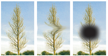
When the macula is not functioning properly, we notice blurry or darkness in the center of our visual field. Initial symptoms include blurring of written words on a page, straight lines appearing distorted, or a black or white spot appearing in the middle of the center of vision.
HOW MANY TYPES OF YELLOW SPOT DISEASE ARE SEEN?
There are two types of macular degeneration (macular degeneration);
Dry type: It occurs with the accumulation of lipid (oily) material under the retinal layers. It progresses more slowly and causes vision loss in the long term.
Wet type: It occurs when damaged capillaries in the patient area progress to the macula. This type can cause sudden vision loss. It is less common than the dry type, but causes 80% vision loss. Especially if the patient has membrane formation in one eye, the other eye is also in danger.
WHAT ARE THE RISK FACTORS?
Surgical removal of the membranes formed in this area or trying to provide vision by shifting the center of healthy parts of the retina is a developing method today.
The earlier this disease is diagnosed and the patient is followed up, the better vision loss can be prevented.
YELLOW SPOT DISEASE TEST (Amsler Grid Test)
With adequate lighting on, put on your reading glasses and stand 20-25 cm away from the screen. stay close.
Close one eye.
Look straight at the dot in the middle with your open eye.
When looking at the point, all the lines around it are straight or distorted,
Check if it is blurry or dark.
Repeat the same process with your other eye.
Part of the test is wavy, blurry or dark
If seen, call your ophthalmologist.
This disease can occur in one in every four people who have a first-degree relative with macular degeneration, usually in people over the age of 60, and women are at higher risk than men of the same age.
In addition, factors such as diabetes, hypertension, smoking, ultraviolet light, light-coloured eyes, arteriosclerosis, heart enlargement and vitamin deficiency are also included in the risk group.
PHOTODYNAMIC THERAPY
In recent years, a different laser treatment, photodynamic therapy (PDT), has been applied by administering a special dye, affecting only the capillaries and not damaging the underlying retinal cells.
After the treatment, the closure of the membrane is checked with angiography. If necessary, PDT is repeated after three months. Up to 60% success rate is achieved with this technique, and at least vision loss is prevented.
WHAT ARE THE CAUSES AND SYMPTOMS?
Diabetes causes damage to capillaries in various parts of the body. Conditions such as pregnancy, hypertension and smoking increase the damage of diabetes to the retina. In the early period, when edema does not occur in the macula, which is our sharp point of vision, the changes caused by diabetes in the eye do not cause any symptoms. These can only be detected on examination. At more advanced levels, vision becomes blurred and sometimes completely lost due to bleeding. Therefore, regular examinations are required at six-month intervals.
WHO IS AT HIGH RISK OF DIABETIC RETINOPATHY?
Diabetic retinopathy can happen to anyone with diabetes. The most important factor is the duration of diabetes. The longer the period, the higher the risk of diabetes-related eye disease. High blood sugar levels and blood fats, hypertension and kidney disease further increase eye disorders due to diabetes.
DIAGNOSIS AND HOW IS DIAGNOSIS DONE?
You must undergo a full examination by an ophthalmologist. Severe retinopathy sometimes causes no symptoms and may respond to treatment. Therefore, diabetic patients should know the risks they face and have their eyes examined regularly. During the examination, the pupils are dilated and the retina is observed painlessly with devices called ophthalmoscopes. If there are symptoms of diabetic retinopathy, a special angiogram is performed and the stage of the disease and treatment scheme are determined.